IRDye® 680LT Goat Anti-Mouse IgG2a
|
926-68051 |
Highly Cross-Adsorbed Goat Anti-Mouse IgG2a (γ2a Chain Specific) Antibody Conjugated to IRDye 680LT |
Applications
IRDye 680LT subclass specific antibodies can be used for a variety of applications, including Western blotting. These antibodies are optimized for use with instruments within the Odyssey Imagers. Two-color detection can be achieved by multiplexing IRDye 680LT subclass specific antibodies with IRDye 800CW subclass specific antibodies. IRDye 680LT subclass specific antibodies are also suitable for immunofluorescent microscopy and other fluorescent imaging applications when using instrumentation with appropriate excitation and detection capabilities.
IRDye 680LT is not intended for in vivo applications.
Visit licorbio.com/support for protocols, example experiments, recommendations, and more technical information.
Storage
- Storage temperature: 4 °C
- Storage conditions: Protect from light and moisture.
-
Shelf life: When stored as recommended, this product is stable for 3 months.
Specifications
-
Fluorophore: IRDye 680LT (MW 1402)
-
Size:0.1 mg
-
Antibody Concentration:1.0 mg/mL(when reconstituted as directed)
-
Form of Antibody: IRDye 680LT purified immunoglobulin conjugate, lyophilized in phosphate buffered saline, pH 7.4. Contains 10 mg/mL BSA (free of IgG and protease) as stabilizer and 0.01% sodium azide as preservative, after reconstitution.
Contains sodium azide.
-
Excitation Wavelength: 679 nm (in PBS)
-
Emission Wavelength: 696 nm (in PBS)
-
Immunogen: Mouse IgG2a paraproteins
Purity and Specificity
Isolation of specific antibodies was accomplished by affinity chromatography on mouse IgG2a covalently linked to agarose. Based on ELISA and flow cytometry, this antibody reacts with the heavy chain of mouse IgG2a. This antibody was tested by Dot Blot and/or solid phase adsorbed for minimal cross-reactivity with mouse IgM, IgG1, IgG2b, IgG3, IgA, pooled human sera, and purified human paraproteins. The conjugate has been specifically tested and qualified for Western blot. Results with your primary antibody may vary and specificity should be confirmed prior to performing two-color detection.
Reconstitute Secondary Antibody
-
Combine each 0.1 mg of secondary antibody with 0.1 mL of sterile distilled water.
-
Gently mix by inverting.
-
Allow the solution to stand at room temperature for at least 30 minutes before use. Protect the solution from light.
If the solution is not completely dissolved after standing at room temperature, centrifuge the solution.
When stored as recommended, this product is stable for 3 months.
Recommended Dilutions
Optimum dilutions will vary and should be determined empirically.
Avoid non-specific background staining on PVDF membranes: If you are making your own antibody diluent, add SDS (final concentration 0.01 – 0.02%) and Tween® 20 (final concentration 0.2%) to the diluent. If using Intercept® T20 Antibody Diluent, only add SDS (final concentration 0.01 – 0.02%).
Recommended Dilutions for Western blotting
| Recommended | Suggested Range |
| 1:20,000 | 1:10,000 – 1:50,000 |
Example Data
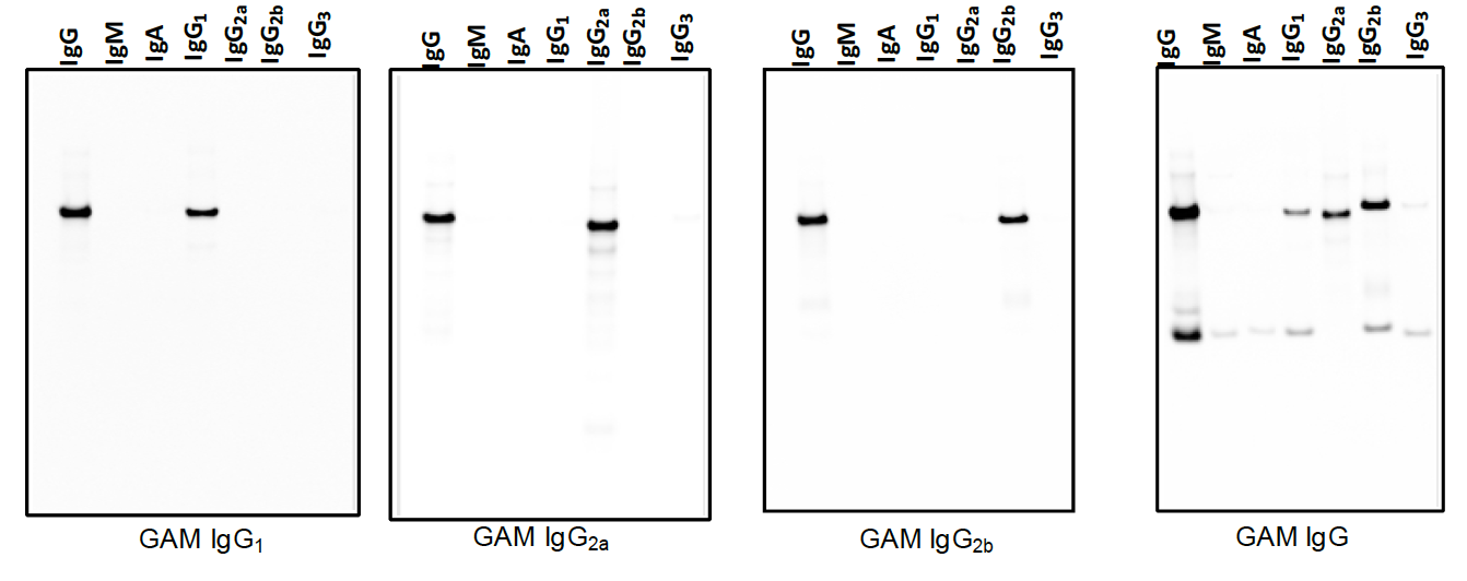
IRDye® subclass specific secondary antibodies react with mouse gamma 1, mouse gamma 2a, or mouse gamma 2b, depending on the secondary antibody used for detection. All other IRDye secondary antibodies are whole IgG (H+L) and react with the heavy (gamma) and light chains. Figure 1 demonstrates the performance of IRDye subclass specific secondary detection compared to IRDye whole IgG for detection of purified mouse subclass antibodies.
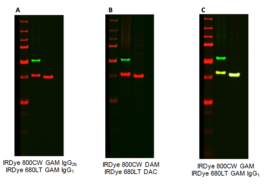
When performing two-color Western blot detection, it is important to choose the IRDye secondary antibodies based on the primary antibodies being used. IRDye subclass specific antibodies allow for two-color detection using primary antibodies derived from the same species (mouse). Figure 2 demonstrates two-color Western blots using subclass specific detection (A) and whole IgG detection (B). IRDye subclass specific secondary antibodies should not be used in combination with IRDye whole IgG secondary antibodies for two-color detection. IRDye whole IgG secondary antibodies and subclass specific secondary antibodies both recognize the gamma chain of the primary antibody, causing detection in both channels (C).