Histone H3 Rabbit Monoclonal Antibody
Specifications (926-42219)
-
Size: 100 µL
-
Storage: -20 °C
-
Species Cross-Reactivity: Human, mouse, rat, monkey
-
Target Molecular Weight: 17 kDa
-
Isotype: Rabbit IgG
-
Specificity/Sensitivity: Detects endogenous levels of total Histone H3 protein (including isoforms H3.1, H3.2, and H3.3) and Histone H3 variant CENP-A. Does not cross-react with other core histones. May cross-react with bovine, chicken, D. melanogaster, hamster, xenopus, and zebrafish.
-
Immunogen: A synthetic peptide that corresponds to the carboxy terminus of the human histone H3 protein
-
Storage Buffer: 10 mM HEPES (pH 7.5), 150 mM NaCl, 100 µg/mL BSA, 50% glycerol, and <0.02% sodium azide
Do not aliquot the antibody.
Contains sodium azide.
-
Tested Application: Western blot, Immunohistochemistry, Immunofluorescence, Flow Cytometry
Recommended Dilution
-
Western Blotting: 1:1000
Applications
The Histone H3 primary antibody can be used as an internal loading control for normalization and is particularly effective when detecting target proteins in nuclear extracts.
The expression of Histone H3, or any housekeeping protein (HKP), should be validated to ensure that its expression does not change under experimental conditions.
Once validated, Histone H3 primary antibodies can be used for the detection of Histone H3 when performing multiplex Western blot detection.
Detect Histone H3 Rabbit Monoclonal Antibody with IRDye® Goat anti-Rabbit or IRDye Donkey anti-Rabbit secondary antibodies.
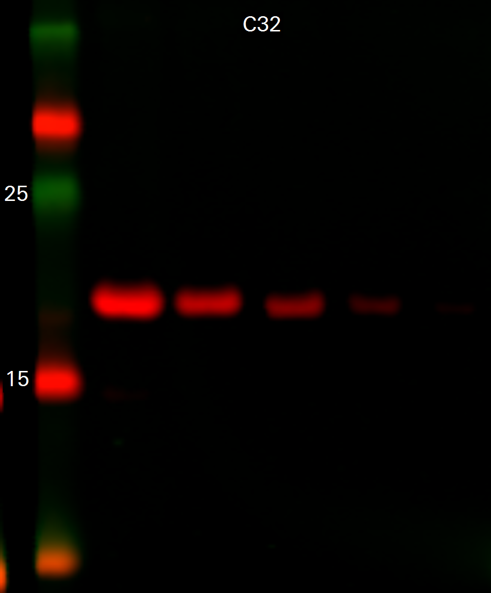
Histone H3 Rabbit Monoclonal Antibody in C32 Lysates
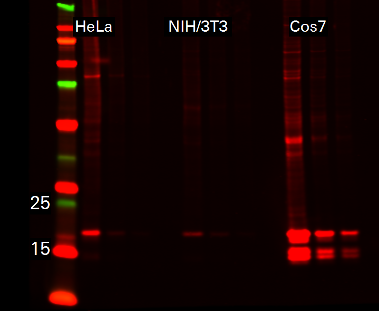
Histone H3 Rabbit Monoclonal Antibody in HeLa, NIH/3T3, and Cos7 Lysates