Host Cell Proteins and Antibody Characterization
Cell culture is often used in the production of biopharmaceuticals. The host cells express not only the drug itself, but also many other proteins called host cell proteins (HCPs). These can impair the efficacy of the active ingredient and potentially be dangerous to humans. Therefore, the cut-off value of HCPs must be qualified and quantified after purification of the product.
1. Sample Preparation
Most commercially available purification kits for 2D electrophoresis are bind-wash-elute columns. These are expensive, cause a lot of plastic waste, and always involve protein loss. In case of many or different impurities, we recommend protein precipitation according to Wessels & Fluegge. This removes all impurities and salts and protects the sample. For precipitation, we offer methanol and chloroform.
Methanol and Chloroform

2. 2D Separation
First Dimension: Isoelectric Focusing on IPG Strips
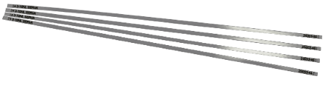
In isoelectric focusing, proteins are separated according to their isoelectric point. For this purpose, gel strips with immobilized pH gradients (IPG strips) are used. According to the separation requirements of your sample and depending on the choice of gel format (mini-midi-large), we offer five different lengths (7–24 cm) with six different pH gradients each. In HCP analysis, 24 cm gels with a pH range of 3–10 are used to obtain the maximum in expanse combined with the whole pH spectrum, eliminating data loss.
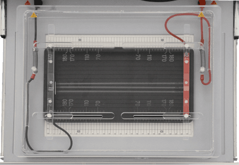
HPE™ Electrode Lid with IPG Tray
Using the HPE™ electrode lid with IPG tray, you can upgrade your HPE BlueHorizon™ to an IPG focusing device.
Second Dimension: SDS PAGE on Blottable Gels
In SDS PAGE, proteins are separated according to their molecular weight. Especially in 2D electrophoresis, the spot sharpness is important for a good evaluation. The ultra-thin constantly cooled horizontal gels increased the spot sharpness manyfold. To prevent the gels from tearing, they are applied on a carrier foil.
The best chamber for these gels is the HPE BlueHorizon. A ceramic cooling plate, indestructible electrodes, and stackability are just three of the many advantages.
Horizontal, film-backed gels have the disadvantage that they are not blottable. In the past, a 2-liter bottle and a fishing line were used to cut the gel off the film. We have developed blottable gels that adhere covalently to the film only at a narrow, defined area. Using a scalpel, this is cut off and the gel can then be peeled off. To avoid tearing the gel, it is recommended to build up the blot upside-down.
The 2D HPE BlotGels are available in three versions for short, medium, and long IPG strips. The backing is non-fluorescent. It is therefore possible to detect the separated sample after electrophoresis and before blot setup with fluorescent dyes or labels.

2D Chemicals Bundle
Our 2D Chemicals Bundle includes all the chemicals you need for HCP and antibody characterization, such as urea, detergent, DTT, and Iodoacetamide.
- Urea, analytical grade (5 kg)
- CHAPS Zwitterionic Detergent (5 g)
- Dithiothreitol (DTT) analytical grade (5 g)
- Iodoacetamide research grade (5 g)
3. Detection Methods
Two methods are most often used to detect.
2D Chemiluminescence Method
The 2D chemiluminescence method uses two gels that ran in parallel on a HPE BlueHorizon double deck. One gel is stained with a colorimetric protein stain and detected. The second gel is blotted, labeled with an anti-CHO primary and a HRP conjugated secondary antibody, and detected. With our SERVA Purple HiSens total stain, you can raise the sensitivity by adding fluorescence as a detection method.

2D DIBE Method
The 2D DIBE method works with one gel. The sample is pre-labelled with SERVA Lightning Sci3, then 2D electrophoresis is performed and blotted. After blotting, the membrane is labelled with an anti-CHO primary and a Sci5 conjugated secondary antibody. Detection is a multiplex approach.

4. Transfer
With the 2D HPE BlotGels, blotting horizontal gels is simple. A small cut with a scalpel and the gel can be peeled off the plastic backing.
BlueBlot™ Semi-Dry Blotter
The easiest way to do this is with the BlueBlot™ Semi-Dry blotter. The completely removable electrodes make the upside-down setup much easier than with a glass plate. After everything has been stacked bubble-free, the sandwich is flipped between the electrodes and placed in the blotter. A specially designed lid holds the blot in place and presses it lightly—no need to weigh down the lid.
OrderBluePower™ 300 BLOT
Since you’re blotting large gels, you also need a power supply with high ampere output. The BluePower™ 300 BLOT provides enough current for a perfect blot result.
Order2D Blot Bundle
Our 2D Blot Bundle will kickstart your HCP and antibody characterization workflow with buffer, large format gels, and nitrocellulose membranes.
- HPE Blotting Kit for large format gels (3 Kits)
- NC 45 Nitrocellulose Membrane 0.45 µm 30 cm x 3 m (1 roll)
- Towbin Buffer for Western Blotting 10x (1 L)
- Intercept® (TBS) Protein-Free Blocking Buffer (500 mL)
5. Detection
To block non-specific binding sites for your antibodies, we recommend either the protein-free blocking reagent BlueBlock PF or Intercept Protein-Free Blocking Buffer. Another advantage is that it can also be used to dilute your HCP primary antibody and anti-IgG-HRP secondary antibody.
BlueBlock PF

Intercept® Protein-Free Blocking Buffer

For chemiluminescence detection, SERVA gives you the choice between standard A/B solution and ready-to-use pre-mixes. The respective SERVALight substrates differ in light intensity and duration. LICORbio also offers WesternSure® Eco and WesternSure Pro ECL Substrate.
WesternSure® Eco ECL Substrate

WesternSure® Pro ECL Substrate

6. Quantification
Odyssey® Imaging Systems
LICORbio’s Odyssey® Imaging Systems are the gold standard for fluorescence imaging and protein quantification. With 6+ logs of dynamic range, high sensitivity and no saturation, and over 85,000 citations in peer-reviewed publications, Odyssey Imagers have become synonymous with reliable quantification for a wide variety of applications.

LICORbio Atlas™ Imaging System
The LICORbio Atlas Imager is a versatile screening platform that delivers quantitative data across a wide range of assays in fluorescence, luminescence, and brightfield. Built on 30 years of trusted LICORbio technology, this instrument delivers unique screening capabilities that help you save time, money, and lab space.

7. Software and Data Analysis
Image Studio™ Software
Quantitative protein data analysis requires reliable, trustworthy software. With over 150,000 downloads worldwide, LICORbio’s Image Studio™ Software is a trusted standard for flexible, reproducible image acquisition and analysis. Its adaptable design provides greater control over analysis parameters in a single package, without manipulating the image. It is optimized to support Odyssey Imaging Systems and the LICORbio Atlas Imager.

Frequently Asked Questions (FAQs)
What are HCPs?
HCPs is an abbreviation for Host Cell Proteins. Host cells, such as CHO cells or E. coli, are used to produce biologicals or pharmaceuticals. Remaining proteins of the HCPs might cause an immune response. Regulatory does want to have evidence of the lowest possible amount after production.
Why does my HCP analysis result show vertical or horizontal streak?
The most crucial impact in 2D electrophoresis and thus HCP analysis is the sample itself. Especially salts and phosphates from the culture media have an impact. Good sample preparation using kits, precipitation, or buffer change can help. Although sample preparation can reduce the protein amount, the loaded concentration should at least have 50-100 µg per strip.
What can be done against a strong background after blotting an HCP sample?
Especially the blotting of HCP samples can cause a strong background due to residues of cells, culture media, or media substances. An overnight washing step after the first blocking step with Intercept Blocking Buffer or any other blocking agent can reduce the background.
What are the differences between HCP analysis using 2D electrophoresis, ELISA, and LC/MS?
2D electrophoresis is highly sensitive and produces the highest resolution. ELISA is only a fast overview technique, and LC/MS works without a specific antibody.